How does blood flow across the placental surface affect fetus size?
Associate Professor Jo James from Waipapa Taumata Rau the University of Auckland will explore how blood flow across the placental surface impacts the birth of dangerously small pēpi
Published on 3 Whiringa-ā-rangi November 2022
One of the most complex blood spaces in the human body, called the intervillous space, develops in pregnancy. It is an expansive vascular space with a tree-like structure involved in placental exchange – where blood flow carries nutrients and oxygen between the whenua and intervillous space – which is critical for growing a healthy fetus. An impaired placental exchange can lead to a condition called fetal growth restriction, when pēpi babies are born dangerously small. This is a significant clinical issue and has no treatment other that preterm delivery. Over 50% of fetal growth restriction cases are not detected prior to delivery, which makes this condition the largest risk factor for stillbirth.

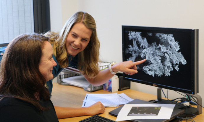
Associate Professors Jo James and Alys Clark (sitting) examine placental cells under the microscope. Photo supplied
A unique cell called the syncytiotrophoblast on the outer surface of the whenua can sense blood flow through the intervillous space. Previous work from the Pregnancy Modelling Group that Associate Professor James co-leads with Associate Professor Alys Clark has suggested this sensing results in a dynamic relationship between blood flow and whenua development across pregnancy. However, there has been little consideration how the interplay between blood flow and whenua tissue architecture evolve over the course of pregnancy.
Associate Professor James and her team have been awarded a Marsden Fund Standard grant to investigate how blood flow across the placental surface changes in fetal growth restriction. They will look at the responses of shear stress on the syncytiotrophoblast cell of the whenua, and the impacts of this cellular stress on whenua development and function. This project will combine cutting-edge bioengineering tools, such as 3D Micro-Computed Tomography imaging, with stem cell tools. The overall goal is to tease apart the relationships between whenua architecture, intervillous space blood flow, and syncytiotrophoblast cell function that are fundamental for human life to develop. These insights are imperative to shed light on the underlying processes of fetal growth restriction and improve the ability to identify at-risk pēpi.